Summary of microbe sampling, sample preparation techniques and dilution, inoculation and culture methods!(2)
1.Inoculation method of sample solution:
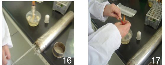
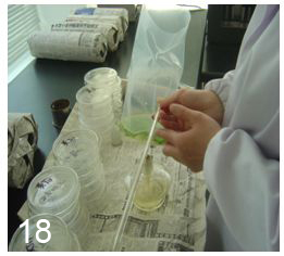
1.1. Pull out the suction tube along the upper wall of the tube, blow and wash the suction ear ball beside the flame, insert the suction ear ball at the end of the suction tube, quickly pass through the flame from the end of the suction tube to the tip, and empty the remaining water in the suction tube, as shown in Figure 16, 17 and 18.
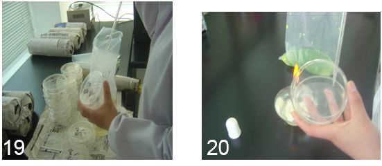
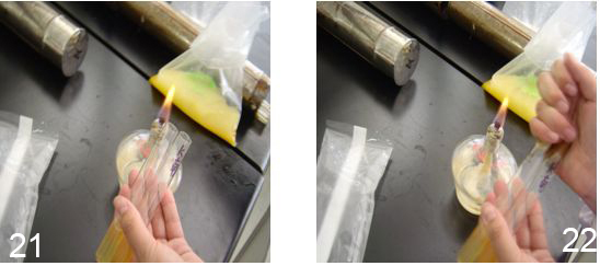
1.2. Insert the straw into the homogenized bag at a depth less than 2.5cm, and accurately absorb the homogenized sample (the tip of the straw should not touch the mouth of the bag). The liquid should be drawn above the required scale, then the pipepipe should be lifted so that the tip of the pipepipe is removed from the liquid level and attached to the bag to adjust the liquid to the required scale. 1ml, 1ml, 1ml, 0.2ml aseptic operation were respectively completely injected into the labeled number of bacteria, coliform group, EC and staphylococcus aureus plate within 2 ~ 4s (EC should be rapidly shaken and uniform after inoculation), figure 19, 20, 21, 22.
1.3. If a sample solution is left for more than 3 min before inoculation, it should be re-homogenized.
1.4. In order to verify the sterility of diluents, media, dishes, pipettes and other utensils, blank control should be made.
2.Pour pour method
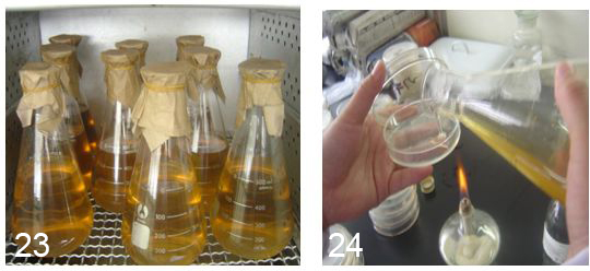
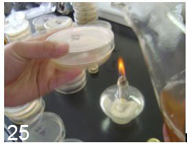
Hold the triangle flask of medium in the right hand (it has been placed in a water bath at a constant temperature of 50-60℃) next to the flame, open the plate into a seam with the left thumb and index finger or middle finger, quickly pour about 15ml of medium, and gently shake the plate after the lid is covered, so that the medium is evenly distributed at the bottom of the plate, and then flat on the table; Alternatively, open the petri dish with the left thumb and index finger on a table near the flame, then inject the medium, and shake well, as shown in Figure 23, 24, 25.
Does note:
A. The medium should not be kept warm for more than 4h in a water bath of 50-60℃.
B. The operation shall be limited to 20min from the beginning of sampling to the subinjection of the medium.
C. The coated plate surface should be dry (insufficient drying is prone to bacterial colony diffusion, condensate water is not conducive to bacterial separation and will lead to bacterial reproduction and distortion of results; Excessive drying will cause the medium to split and cannot be used; If the drying is suitable, the sample liquid will be absorbed completely and quickly.
D. Bacteria are easy to adsorb on the surface of glassware, so the sample liquid in the plate should be fully mixed with the AGAR medium as soon as possible after the bacterial liquid is injected into the plate, otherwise the bacteria will not be easily dispersed. The mixing method is to tilt and rotate the plates to fully mix, but care should be taken not to allow the medium to overflow from the plate and not adhere to the plate walls and cover.
3. Cultivation
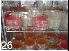
3.1. Invert the plates into the constant temperature incubator (a maximum of 6 plates should be stacked per stack, and there should be space between the plates for air circulation, so that the temperature of the culture can reach the same as the temperature of the incubator as soon as possible), and the culture should be carried out according to the specified time and temperature. The incubator should maintain a certain humidity, and the weight loss of AGAR medium cultured for 48 hours should not exceed 15%, as shown in Figure 26.
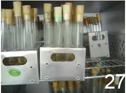
3.2. The inclined plane, the high-rise inclined plane and the liquid medium are placed on the test tube rack into the constant temperature incubator, and the culture is carried out according to the specified time and temperature, as shown in Figure 27.
4. Count determination and matters needing attention
4.1. Because there are so many kinds of bacteria, they are very different. When counting, the transmitted light is usually used to observe the back or front of the plate (colony counter) carefully. If necessary, inspect with a magnifying glass to avoid missing colonies, especially on the edges of the dishes.
4.2. Refer to SN/T 0168-2015 for counting method.
4.3. In a low dilution petri dish, it is difficult to distinguish between microbial colonies and food debris. Generally, food debris is not shiny and not smooth enough to identify.
4.4. In order to avoid the small particles in the food or the impurities in the culture group and the bacterial colony confusion, it is difficult to distinguish, can be simultaneously used as a diluent and AGAR culture group mixed plate, without culture, and placed at 4℃ environment, in order to count for control observation.
4.5. When necessary, in order to prevent food particles from being confused with colonies, TTC (100ml plus 1mL 0.5%TTC) of triphenyltetrazole chloride can be added to plate counting AGAR. Colonies will be red after culture and easy to distinguish.
4.6. In the fermentation test, if the gas production of the fermentation tube is more than that of the fermentation tube, it can be filled with gas. If the gas production of the fermentation tube is less than that of the small rice grain, it can produce bubbles smaller than the small rice grain, or there are small bubbles rising slowly along the wall of the fermentation acid, it should be further tested. If in doubt about the fermentation which produces acid but not gas, the tube can be gently touched by hand. If there are bubbles floating along the wall of the tube, as is the case with carbon dioxide bubbles in beer, the possibility of gas production should be considered and further testing should be carried out. If it’s just a shaking bubble, it’s negative.
Post time:2024-08-01
