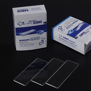
The making of microscope slides is a process that consists of several steps and techniques, which may vary depending on the aim of the research and type of sample.
Choosing the right slide and cover slip:
To begin with, one has to select the appropriate slide as well as cover slip. When it comes to observing living cells, there could be need for specific formats of slides such as slide support structures for observing living cells.
Sample preparation:
For each sample type, there are certain procedures applied in preparing them. As an example, in case you have small particles that are randomly distributed then this can be done using Moore method whereby a sink is filled with particles suspension after which a slide or cover slip is put at the base then it is stirred to allow particles to settle on the slide. Before slides can be made, cells meant for chromosome preparation should be fixed overnight using fixative.
Mounting of slides and coverslips:
The sample that has been processed should be placed onto a slide and lightly covered by fitting coverslip over it. Throughout this process it must not form air bubbles on it. To ensure even distribution of sample during dropper device can be used while bubble formation may get reduced by changing angles.
Drying and Fixing:
Drying and fixing are essential aspects when handling specimens on slides and coverslips. This step helps maintain specimen’s integrity hence avoiding movements within its cells. Quality final result may depend on drying time as well as environmental conditions like humidity levels in relation to room temperature.
Cleaning and Inspection:
After all these steps have been accomplished, one needs to examine the slide closely so as to know if any contaminant occurred during experimentation around it or damage was caused then either extra cleaning can take place or re-making.
Use of Virtual Slide Technology:
Additionally virtual slides technology may also come into play due to advancing technology. Through computer generated images, this technology enables viewing and doing investigations without any actual slides.
Putting everything together, it is a very precise and delicate process to make microscope slides. Each step must be executed carefully in order for the microscopy to give high-quality observations.
References
1. D. Romer, K. Yearsley et al. “Using a modified standard microscope to generate virtual slides..” The Anatomical Record Part B The New Anatomist (2003).91-7.
2. D. Lazarus. “An Improved Cover-Slip Holder for Preparing Microslides of Randomly Distributed Particles: RESEARCH METHOD PAPER.” (1994).
3. P. Miles and H. M. Dott. “The construction of a membrane slide support for the observation of living cells.” Journal of Microscopy (1979).
4 M.R. Ahlada Rao, R. Victor et al. “Slide Making and Prototype of a Dropper Device for Chromosomal Preparations.” (2005). 213 – 216.
Post time:2024-08-02


