God operation: blood wet film 2 seconds to distinguish true and false thrombocytopenia!
Special statement: This operation does not comply with the re-examination rules of routine blood samples, but it has certain significance for the rapid identification of EDTA-dependent pseudothrombocytopenia, which is only for your colleagues' reference.
EDTA-dependent pseudothrombocytopenia (EDTA-PTCP) is caused by platelet aggregation after EDTA is used to anticoagulate whole blood.
Not much nonsense, the pictures and texts teach you how to make wet slices of blood samples and how to identify platelets in the wet slices.
Item preparation: microscope, glass slide, pipette, specimen to be inspected.
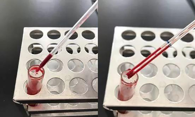
1.The sample to be inspected is gently inverted and mixed, then the lid is opened, and a small amount of the sample is sucked with a disposable pipette or a micro pipette (see the figure below):
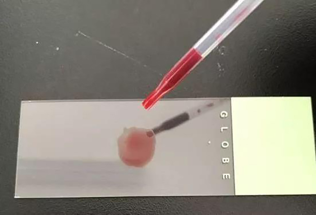
2.Drop the blood onto the glass slide and spread it out with a pipette (see the picture below)
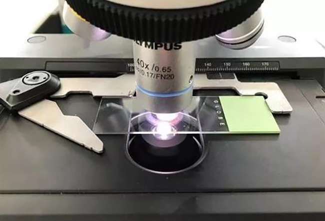
3.Place the glass slide on the microscope, first use a low-power lens (yellow lens) to find the object image, then switch to a high-power lens (blue lens), select the edge of the blood drop that is not dry and suitable for observation (see the figure below) ):
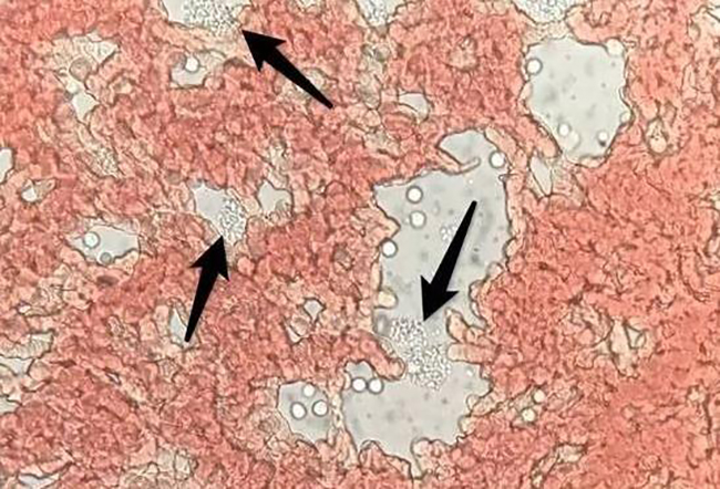
What do the platelets look like in wet tablets?
The following picture shows a peripheral blood sample of a patient with EDTA-dependent pseudothrombocytopenia, showing the distribution of platelets in piles (pointed by the arrow):
The following picture shows a peripheral blood specimen of a patient with aplastic anemia, with hypothrombocytopenia, and almost no platelets are seen in the picture:
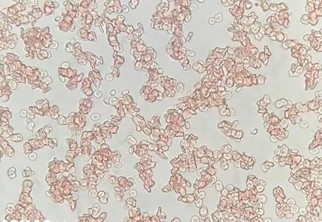
The following picture is a normal human blood sample, showing the scattered distribution of individual platelets:
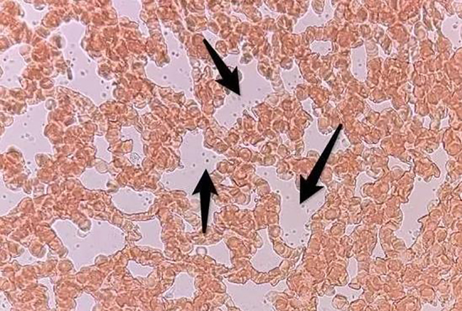
Summary: This method is simple, fast and low-cost. It is suitable for quickly identifying EDTA-dependent platelet aggregation. Accumulating experience will help improve work efficiency. Especially for the case where the basic hospital does not have Wright's (Ruiji's stain), it is very helpful for quickly identifying the true and false reduction of the patient's platelet!
Reminder: In the case of Wright (Rigi's Stain), blood smear staining microscopy should be carried out in strict accordance with the blood routine re-examination rules to avoid missed diagnosis of blood diseases! !
Post time:2024-08-01


