![]()

1.Introduction
In the realm of complex microscopy, where precision meets exploration, even the tiniest aspects can be extremely important. Among many other instruments available to a scientist, microscope slides are an essential but silent partner. Not all slides are made equal. Enter the frosted end microscope slide, an unobtrusive advance that does more than provide a place to hold your sample. This article explores frosted microscope slides: what they are, why they matter, and how they enhance your microscopy work.
2.What Are Frosted Microscope Slides?
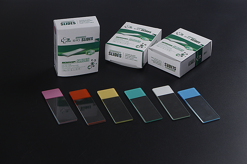
Frosted microscope slides are essentially glass slides like their clear counterparts but with one major difference – some part of their surface is intentionally roughened or “frosted.” This frosted area generally takes up one end of the slide where it is possible to write or etch labels without smudging or erasing them off accidentally. For example in labs these show the dual purpose of specimen holding and easy labeling.

● Why the Frost? The Purpose Behind the Design
This may seem like just any other feature but actually serves a critical purpose. In science work, clarity and organization reign supreme. The ability to label directly on a slide using pencil or special ink that does not erase easily allows meticulous documentation and reduces misidentification risks which is an important consideration for research work and diagnostics as well. These labels will still be readable even after putting them into contact with chemicals and stains used in microscopy thanks to its frosted surface.
● The Benefits of a Frosted Surface: From Labeling to Light Diffusion
Apart from being handy for labeling purposes, there are numerous advantages associated with frosted microscope slides too. The rough texture helps diffuse light, which can be very useful in reducing glare while boosting contrast when looking through microscopes. This property becomes particularly useful when dealing with transparent or low-contrast specimens. Furthermore, this type of glass resists many chemicals meaning that its label will remain intact even under the process of staining thus making it a popular choice in various laboratory settings.
● Comparing Frosted and Non-Frosted Slides
On comparison, frosted and non-frosted slides have each their own benefits. Non-frosted slides are transparent all through and are suitable for situations that require maximum light transmission. However, they do not have the convenience of labeling, which can cause mix-ups in identifying specimens. While slightly diminishing light transmission in the frosted region, frosted slides bring about an important benefit including ease and durability in labeling. For many people this is a compromise worth taking particularly when dealing with clinical or educational environments where correct identification of samples is very crucial.

3.Practical Applications:
● When and Why to Use Frosted Microscope Slides
Frosted microscope slides are made for times when accurate labeling matters most. These are commonly found in pathology labs, schools as well as research institutions. Being able to write directly on them has its huge advantages especially when there are a number of specimens involved. In long-term studies when samples may be revisited many times over, these come handy since they will still be legible even after some time has elapsed and therefore serve their intended purpose.
● Perfect for Staining: Enhancing Visibility and Contrast
For stained microscope slides, one characteristic stands out. Many staining techniques can be used on frosted microscope slides without causing interference. High contrast images can be obtained due to the fact that the frosted surface does not hinder staining. For instance, when using hematoxylin and eosin for tissue samples or Gram staining for bacteria, the use of a frosted slide will keep labels intact while enhancing visuality of specimens.
● Ideal for Teaching and Research: Making Notes Directly on the Slide
Frosted glass microscope slides are convenient in that they allow educators and researchers to take notes directly onto them. This is especially useful in teaching contexts whereby learners can label parts of the specimen as an aspect of their learning process. At the same time, it provides researchers with a way of documenting observations more fully without needing another document so that their work becomes more efficient.
● Frosted Slides in Pathology:Why Precision Matters
The precision provided by using frosted microscope slides is invaluable when it comes to pathology where correct identification of tissues may mean life or death situations. They usually deal with multiple slides at any given time and therefore being able to clearly mark each slide permanently is vital if mix-ups are to be avoided. The frosting ensures that even when touched repeatedly, exposed to substances they still maintain their clarity thus avoiding ambiguity.
● Veterinary and Clinical Uses: Ensuring Accurate Sample Identification
Veterinary clinics and clinical laboratories also benefit greatly from the use of frosted microscope slides. In these settings, where a wide variety of samples are handled daily, there is high risk of cross-contamination or misidentification rates occurring; consequently they require such frosted microscopy slides which help them maintain proper record keeping so that each sample can be identified accurately before processing.

4.Cleaning Tips:
● Maintaining Clarity and Integrity
Cleaning frosted microscope slides requires careful handling because any wrong move could ruin them. Glass needs to be cleaned using a non-abrasive agent so as to avoid scratching the frosted surface and impairing its transparency. For this purpose, it is advisable to take a soft piece of fabric that has been moistened with mild soap or special glass cleaner. This means that thorough cleaning should be done with distilled water only so that residue does not interfere with the microscopic observations. Finally, it is important to dry it off gently using a lint-free cloth in order to prevent leaving strands stuck on the slide.
5.Storage Solutions:
● Protecting the Frosted Surface from Wear
The proper storage mechanism goes a long way in ensuring that the frosted microscope slides are preserved well and remain intact. It is advisable for one to keep them in an environment which is free from dirt and dampness, preferably settling for a slide box having individual compartments that will prevent one from rubbing against another while inside such container. Keep them away from dusts as well as moisture since these destroy both frosted and clear surfaces over time.
6.What to Look for in Quality Frosted Slides
When buying frosted microscope slides, always consider whether you have purchased too rough or excessively smooth ones since they are prone to frosting inconsistencies. The glass used should be of high optical quality without bubbles or streaks present because these may otherwise affect microscopic observations’ results. In addition, the thickness and edge finish of the slide should also be accounted for; they can impact on how durable it is and how good images come out after use.
7.Where to buy frosted microscope slides
When buying frosted microscope slides, purchase from trusted suppliers for quality. For instances, Thermo Fisher Scientific, Globe Scientific or VWR make reliable products that last. These dealers also mean the slides are standardized thus constant performance in your lab work.
8.Conclusion
Frosted microscope slides may seem like a small detail in the grand scheme of scientific research, but they play a crucial role in ensuring precision and accuracy. These slides are an important part of any laboratory, ranging from their labeling convenience to their enhancement of microscopic observations. Frosted microscope slides give you the best combination of practicality and functionality whether you are involved in teaching, research or clinical diagnostics.
9.Reference
1.Doe, J., & Smith, R. (1956). The preparation of frozen-dried tissue for electron microscopy. Journal of Cell Biology, 2(4), 37-45. https://rupress.org/jcb/article-abstract/2/4/37/1464/THE-PREPARATION-OF-FROZEN-DRIED-TISSUE-FOR?redirectedFrom=fulltext
2.Brown, C., & Taylor, D. (2014). Title of the book chapter. In J. Doe (Ed.), Title of the book (pp. 123-145). Academic Press. https://www.sciencedirect.com/science/article/abs/pii/B9780124200678000143?via%3Dihub
Post time:2024-08-15


